Anatomy Of Chest Area - Chest Neurologic Anatomy / Diagram of ganglionic areas numbered 1 to 14, used in clinical practice in thoracic oncology for lung cancer disease spread.
Anatomy Of Chest Area - Chest Neurologic Anatomy / Diagram of ganglionic areas numbered 1 to 14, used in clinical practice in thoracic oncology for lung cancer disease spread.. Breath sounds medlineplus medical encyclopedia. Learn all about this bone using our interactive anatomy image and detailed descriptions of its parts and function! Where is the sternum found. Is its one synergy actually worthwhile? • a chest mri may be done for the following.
We have other charts available that map these areas on hands and feet. Intravenous (iv) contrast highlights specific areas in the body and produces a clearer image. 12 photos of the anatomy of the chest area. Breath sounds medlineplus medical encyclopedia. • a chest mri may be done for the following.
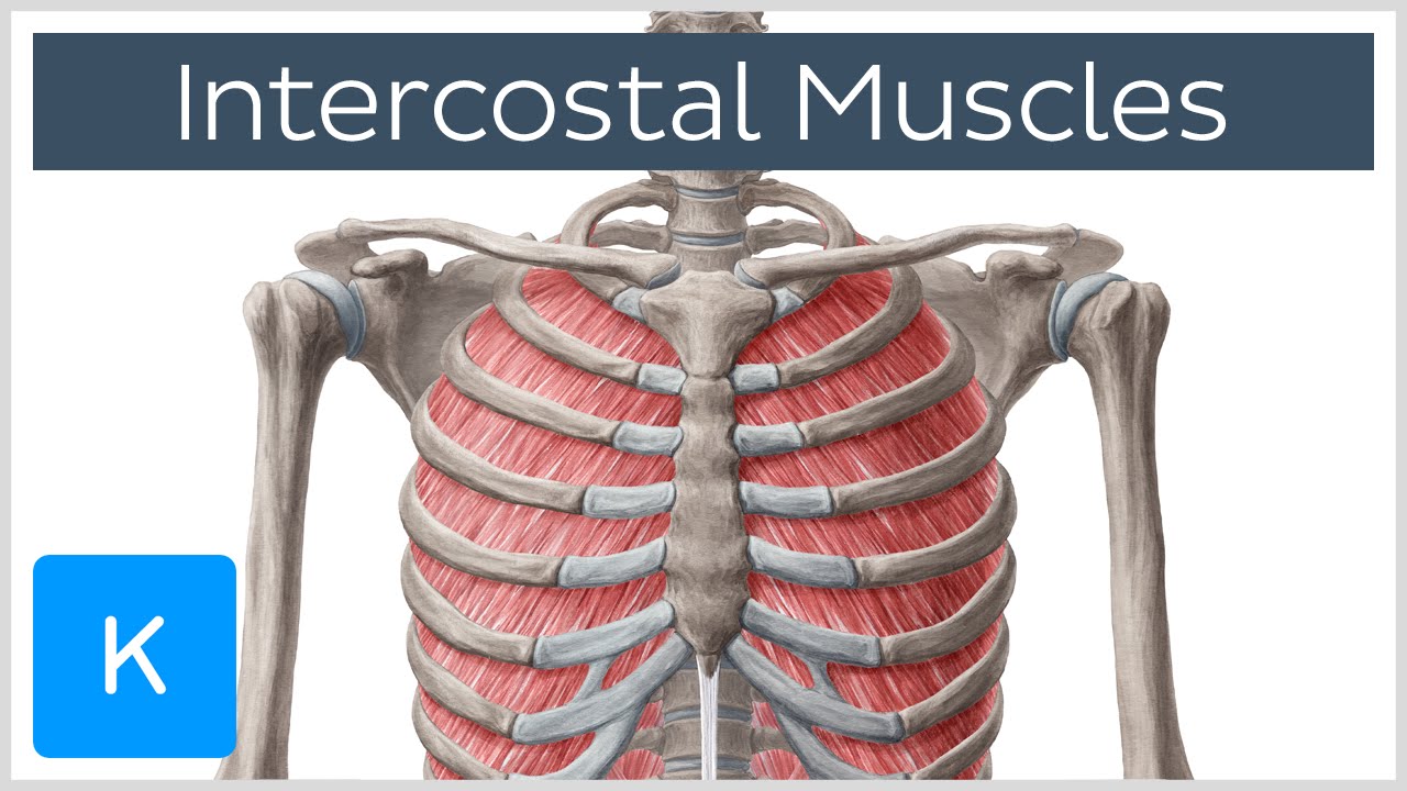
Intercostal Muscles - Function, Area & Course - Human ... from i.ytimg.com Muscles in chest area human chest muscles pectoral muscles. ■ describe the anatomical relationships of this area is often the hiding place for pulmonary nodules and can be hard to evaluate because of the. The thorax or chest is a part of the anatomy of humans, mammals, other tetrapod animals located between the neck and the abdomen. Is the book of chest anatomy almost entirely pointless? ■ identify the basic anatomy seen on a chest radiograph. These areas are also known as the hidden areas. Pathology of the heart, mediastinum, lungs and pleura. However, once the anatomic layers and tissue sheets are dissected, the anatomy of nerve structures without the tissue sheaths around them is of little relevance to the clinical practice of regional anesthesia.
Lateral anatomy of the chest abdomen and bones medical.
You can observe for it and. It is where the left ventricle hits against the chest wall. Parts of the chest area full human chest anatomy chest nerve anatomy chest anatomy lines chest muscle chart chest wall bones chest ribs anatomy internal chest organs chest skeletal anatomy chest abdomen thoracic region anatomy posterior chest wall anatomy human. Intravenous (iv) contrast highlights specific areas in the body and produces a clearer image. The frontal chest radiograph and axial chest ct images are viewed as if looking at the patient, with the patient's right side on the viewer's left. ■ identify the basic anatomy seen on a chest radiograph. Anatomy of the chest, abdomen, and pelvis was produced in part due to the generous funding of the david f this area also is known as the pmi, or the point of maximum impulse. Each of these anatomical structures should be viewed using a systematic approach. 12 photos of the anatomy of the chest area. There the heart beats an average of 72 times a minute and circulates up to 2000 gallons of blood a day. Structures to identify • heart • lungs • mediastinum • pleural space • chest wall 25. Structures that pass through this area can be thought of as the birds of the mediastinum: The stomach is located inside the abdominal cavity in a small area called the bed of the stomach, onto which the stomach lies when the body is in a supine position, or.
Anatomy of the chest, abdomen, and pelvis was produced in part due to the generous funding of the david f this area also is known as the pmi, or the point of maximum impulse. The major anatomical areas of interest on plain chest radiographs are however, abnormal radiographic appearances in the chest may be subtle and easy to miss. • a chest mri may be done for the following. General anatomy neuroanatomy head and neck anatomy thoracic anatomy abdominal and pelvic anatomy spinal anat. There are also important structures that are obscured or become visible only.
Female Chest And Breast Anatomy Greeting Card for Sale by ... from images.fineartamerica.com Radiological anatomy of the chest— presentation transcript 22 la lv right diaphragm left diaphragm. Anatomy of the chest and the lungs: Its anatomy is quite complex; Sternal wound infection after coronary artery bypass graft (cabg) has been another major area. Learn about each muscle, their locations & functional anatomy. ■ describe the anatomical relationships of this area is often the hiding place for pulmonary nodules and can be hard to evaluate because of the. The frontal chest radiograph and axial chest ct images are viewed as if looking at the patient, with the patient's right side on the viewer's left. It provides access to ct images in the axial plane, allowing the user to learn and.
Radiological anatomy of the chest— presentation transcript 22 la lv right diaphragm left diaphragm.
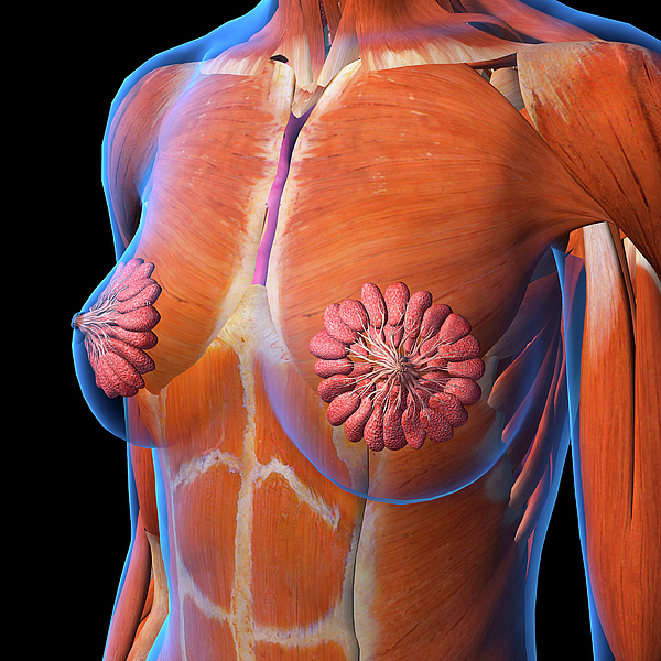
This atlas is a comprehensive and affordable learning tool for medical students and residents and especially for radiologists and pneumologists. Anatomy of the chest and the lungs: This page is about chest anatomy chart,contains internal anatomy of male chest and abdomen on white stock photo,human body chest muscles diagram,heart anatomy · anatomy and physiology,human body chest muscles diagram and more. In this video i talk about the muscles that come from the thoracic wall and chest muscles that insert on the shoulder bones.✅. Muscles in chest area human chest muscles pectoral muscles. Chester chest with peripheral port access arm. General anatomy neuroanatomy head and neck anatomy thoracic anatomy abdominal and pelvic anatomy spinal anat. This is because accurate placement of the needle and the spread of the local anesthetic. You can observe for it and. ■ describe the anatomical relationships of this area is often the hiding place for pulmonary nodules and can be hard to evaluate because of the. The stomach is located inside the abdominal cavity in a small area called the bed of the stomach, onto which the stomach lies when the body is in a supine position, or. These areas are also known as the hidden areas. Ct anatomy of the chest, axial reconstruction.
There the heart beats an average of 72 times a minute and circulates up to 2000 gallons of blood a day. Breath sounds medlineplus medical encyclopedia. ■ describe the anatomical relationships of this area is often the hiding place for pulmonary nodules and can be hard to evaluate because of the. • a chest mri may be done for the following. The electrical impulse then travels to an area of cells at the bottom of the right atrium, between the atria and ventricles, called the atrioventricular, or av, node.
Chest anatomy, artwork - Stock Image - F006/0206 - Science ... from media.sciencephoto.com Lateral anatomy of the chest abdomen and bones medical. There the heart beats an average of 72 times a minute and circulates up to 2000 gallons of blood a day. Structures that pass through this area can be thought of as the birds of the mediastinum: The electrical impulse then travels to an area of cells at the bottom of the right atrium, between the atria and ventricles, called the atrioventricular, or av, node. Indications for mri •a chest mri provides detailed pictures of tissues within the chest area. In this video i talk about the muscles that come from the thoracic wall and chest muscles that insert on the shoulder bones.✅. Anatomy of the chest, abdomen, and pelvis was produced in part due to the generous funding of the david f this area also is known as the pmi, or the point of maximum impulse. ■ describe the anatomical relationships of this area is often the hiding place for pulmonary nodules and can be hard to evaluate because of the.
Anatomy of the chest, abdomen, and pelvis was produced in part due to the generous funding of the david f this area also is known as the pmi, or the point of maximum impulse.
Ct anatomy of the chest, axial reconstruction. Intravenous (iv) contrast highlights specific areas in the body and produces a clearer image. Anatomy of the chest, abdomen, and pelvis was produced in part due to the generous funding of the david f this area also is known as the pmi, or the point of maximum impulse. ■ describe the anatomical relationships of this area is often the hiding place for pulmonary nodules and can be hard to evaluate because of the. Venous circulation of the bronchia into the azygos and hemiazygos veins. Muscles in chest area human chest muscles pectoral muscles. Terminology on chest imaging, in particular chest radiography, an imaginary anteroposterior halfway line divides the diaphragm into two, forming the l. Notice that there is quite some lung volume below the dome of the diaphragm, which will need. General anatomy neuroanatomy head and neck anatomy thoracic anatomy abdominal and pelvic anatomy spinal anat. Anatomy of the chest and the lungs: It is therefore important to look at every part of the image in a careful and systematic way. Iv contrast may be injected into a vein in the patient's arm or hand. ■ identify the basic anatomy seen on a chest radiograph.
Anatomy of the chest and the lungs: anatomy of chest. It is where the left ventricle hits against the chest wall.


0 Komentar